What Causes Red Blood Cells To Stick Together Rouleau Live Blood Analysis Training Course
. Red Blood ...")
Red Blood Cells In The Rouleau Formation, Coloured Scanning Electron Micrograph (SEM). Red Blood ...
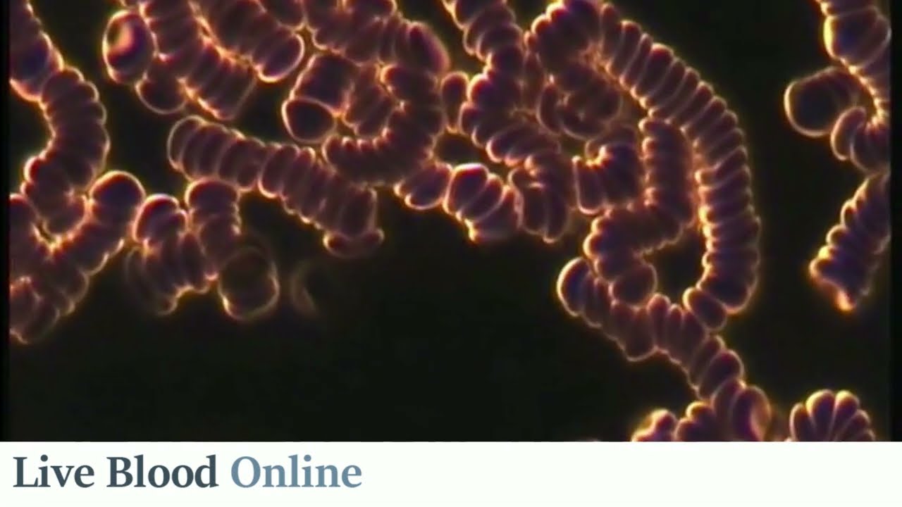
Red Blood Cells In The Rouleau Formation, Coloured Scanning Electron Micrograph (SEM). Red Blood ... The stickiness of red blood cells (rbcs) is due to the appearance of increased serum proteins, often the acute phase proteins such as fibrinogen and immunoglobulins. When an inflammatory process is present, the increase in fibrinogen, α globulins, and/or β globulins in the blood causes red blood cells to stick to each other by interacting with the sialic acid on the rbc surface. the red cells form stacks called ‘rouleau’ that settle faster.
. Red Blood Cells ...")
Red Blood Cells In The Rouleau Formation, Scanning Electron Micrograph (SEM). Red Blood Cells ...
Red Blood Cells In The Rouleau Formation, Scanning Electron Micrograph (SEM). Red Blood Cells ... In pathological states, the increase of plasma proteins (e.g. fibrinogen, globulins) will coat the red blood cells and cause them to become “sticky” and result in rouleaux formation. 1,4. The stacking of cells (rouleaux formation) facilitates the rate of red cell sedimentation, a phenomenon that may be seen on a peripheral smear. the appearance of rouleaux may be artificially caused by a poor preparation of the smear or by viewing the slide in a thickened area. Without considering the possibility of error in sample preparation, a high protein concentration in the blood causes the red blood cells to clump together. the presence of high amounts of the plasma proteins increases the erythrocyte sedimentation rate (esr). Circulating red cells can form cup like structures, allowing them stack into long columns – a process that is promoted and stabilised by large protein molecules present in plasma.
. Red Blood Cells ...")
Red Blood Cells In The Rouleau Formation, Scanning Electron Micrograph (SEM). Red Blood Cells ...
Red Blood Cells In The Rouleau Formation, Scanning Electron Micrograph (SEM). Red Blood Cells ... Without considering the possibility of error in sample preparation, a high protein concentration in the blood causes the red blood cells to clump together. the presence of high amounts of the plasma proteins increases the erythrocyte sedimentation rate (esr). Circulating red cells can form cup like structures, allowing them stack into long columns – a process that is promoted and stabilised by large protein molecules present in plasma. The stickiness of red blood cells (rbcs) is due to the appearance of increased serum proteins, often the acute phase proteins such as fibrinogen and immunoglobulins. It tends to disturb the membranes of the red blood cells so the cells stick together. if you watch this in live blood, you see the white blood cells cleaning the surfaces of the red blood cells. Human erythrocytes in whole blood have a natural tendency to ad here together to form aggregates with characteristic morphology known as rouleaux. the ultimate cause of this phenomenon is the presence in the plasma of large plasma proteins, especially fibrinogen. I spent the last three months learning to do live blood microscopy including bright field and dark field microscopy to detect hydrogels and abnormalities in blood cells caused by graphene oxide and hydrogels from the covid jabs and the environment.
. Red Blood ...")
Red Blood Cells In The Rouleau Formation, Coloured Scanning Electron Micrograph (SEM). Red Blood ...
Red Blood Cells In The Rouleau Formation, Coloured Scanning Electron Micrograph (SEM). Red Blood ... The stickiness of red blood cells (rbcs) is due to the appearance of increased serum proteins, often the acute phase proteins such as fibrinogen and immunoglobulins. It tends to disturb the membranes of the red blood cells so the cells stick together. if you watch this in live blood, you see the white blood cells cleaning the surfaces of the red blood cells. Human erythrocytes in whole blood have a natural tendency to ad here together to form aggregates with characteristic morphology known as rouleaux. the ultimate cause of this phenomenon is the presence in the plasma of large plasma proteins, especially fibrinogen. I spent the last three months learning to do live blood microscopy including bright field and dark field microscopy to detect hydrogels and abnormalities in blood cells caused by graphene oxide and hydrogels from the covid jabs and the environment.
What Causes Red Blood Cells to Stick Together (rouleau) - Live Blood Analysis Training Course
What Causes Red Blood Cells to Stick Together (rouleau) - Live Blood Analysis Training Course
Related image with what causes red blood cells to stick together rouleau live blood analysis training course
. Red Blood ...")
. Red Blood Cells ...")
. Red Blood Cells ...")
. Red Blood ...")
. Red Blood ...")
. Red Blood ...")


Related image with what causes red blood cells to stick together rouleau live blood analysis training course
 - Live Blood Analysis Training Course")


About "What Causes Red Blood Cells To Stick Together Rouleau Live Blood Analysis Training Course"
Comments are closed.